
Figure 4.
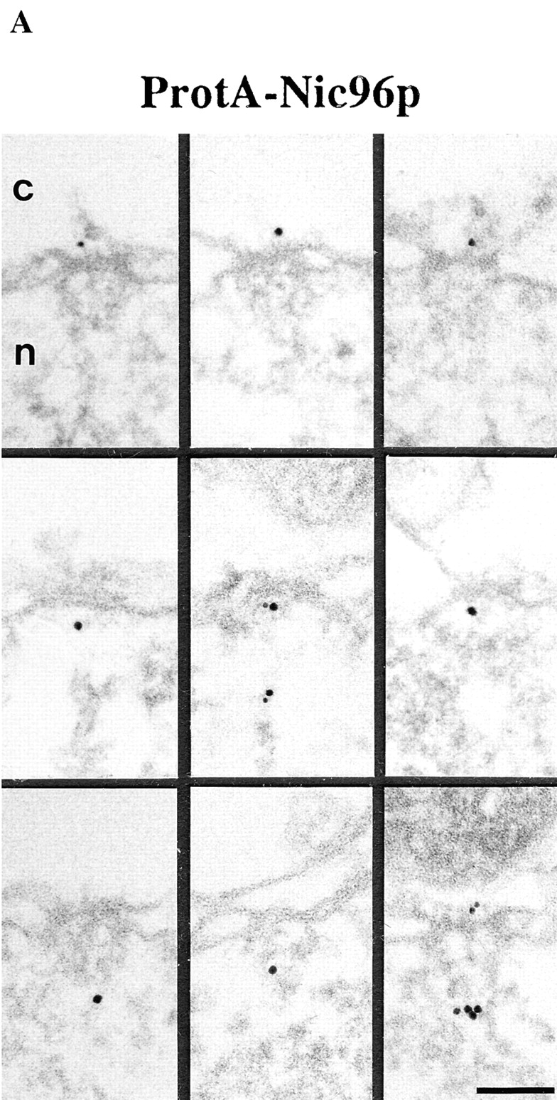

Localization of Nic96p in the ProtA–Nic96p strain. (A) Gallery of selected NPC cross sections showing labeling with the anti–protein A antibody conjugated to 8-nm colloidal gold in Triton X-100–extracted spheroplasts form ProtA–Nic96p cells. Similar to Nsp1p (see Fig. 3), Nic96p is localized at the cytoplasmic (top) and the nuclear periphery (middle) of the central gated channel, and at the terminal ring of the nuclear baskets (bottom). c, cytoplasm; n, nucleus. (B) Quantitation of the gold particle distribution associated with NPCs in the ProtA–Nic96p strain. 80 gold particles were scored. Bar, 100 nm.