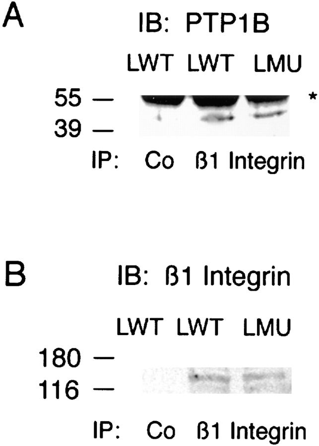
Figure 10.
PTP1B coimmunoprecipitation with β1-integrin. (A) LWT and LMU cells were lysed in nonionic detergent and immunoprecipitated with a rat anti–β1 antibody or normal rat IgG (Co). After separation by SDS-PAGE and transfer to PVDF membranes, the immunoprecipitates were reacted with a rabbit anti– PTP1B and developed with HRP-conjugated anti–rabbit antibody. (B) The membranes were stripped and reblotted with anti–β1-integrin antibody. An asterisk to the right indicates the migration of IgG heavy chain. Numbers at the left indicate the position of molecular mass markers in kilodaltons.

