
Figure 6.
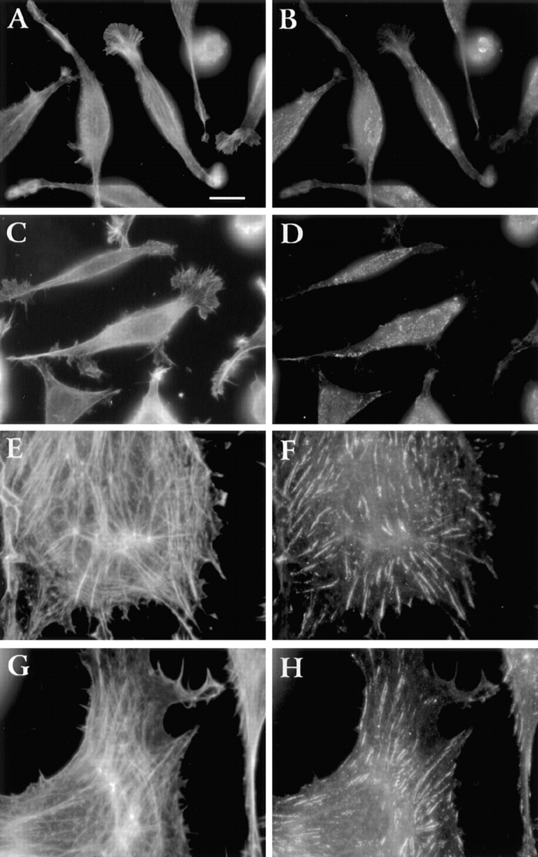
Distribution of β1-integrin and actin in cells cultured for 2 d. LMU (A–D), LWT (E and F), and LP (G and H) cells in DME/FBS were plated on fibronectin-coated coverslips. After 2 d, cells were fixed, permeabilized, and double stained with rat anti– β1 mAb/FITC-goat anti–rat IgG (B, D, F, and H) and TRITC-phalloidin (A, C, E, and G). Bar, 10 μm.