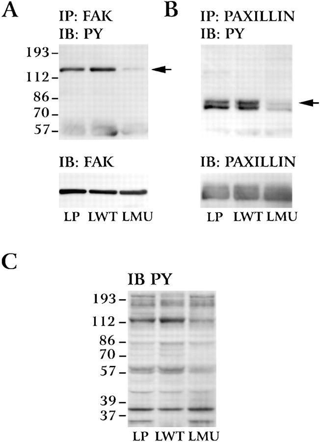
Figure 8.
Phosphotyrosine levels of FAK and paxillin in LP, LWT, and LMU cells. LP, LWT, and LMU cells were plated on fibronectin for 30 min, lysed (see Materials and Methods), and immunoprecipitated (IP) with (A) anti-FAK mAb or (B) antipaxillin mAb and immunoblotted (IB) with antiphosphotyrosine (PY) antibody. Lower panels show stripped membranes reblotted with the same antibodies used for immunoprecipitation. (C) Phosphotyrosine staining of total proteins in Triton X-100 lysates. LP, LWT, and LMU cells plated on fibronectin for 30 min were lysed as described in Materials and Methods. Numbers at the left indicate the position of molecular mass markers in kilodaltons.

