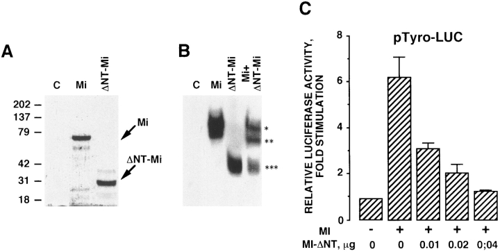
Figure 7.
Mi-ΔNT, a form of microphthalmia lacking the NH2-terminal transactivation domain, exerts dominant-negative effects on the wild-type microphthalmia. (A) Microphthalmia and Mi-ΔNT were in vitro translated in the presence of [35S]methionine, and then analyzed by gel electrophoresis. Molecular masses, indicated on the left, are expressed in kD. (B) For gel shift assay, 2 μl of in vitro–translated microphthalmia, Mi-ΔNT, or microphthalmia plus Mi-ΔNT were incubated with labeled tyrosinase M-box. *, Wild-type microphthalmia homodimer; ***, Mi-ΔNT homodimer; and **, microphthalmia plus Mi-ΔNT heterodimer. Autoradiogram was exposed overnight at −80°C. (C) B16 cells were transfected with 0.4 μg of pTyro, 0.01 μg of pCDNA3 encoding microphthalmia, 0.05 μg of pCMVβGAL, and different amounts of pCDNA3 encoding Mi-ΔNT. After 24 h, luciferase activity was normalized by the β-galactosidase activity and the results were expressed as fold stimulation of the basal luciferase activity from unstimulated cells. Data are means ± SE of five experiments performed in triplicate.

