
Figure 3.
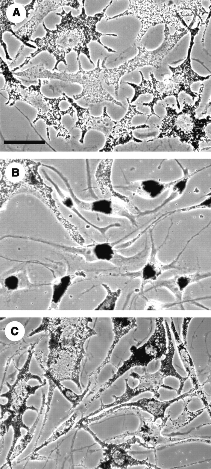
Phase-contrast images of nontransfected cells (A), and cells transfected with the PKA inhibitor plasmid (B), or with the plasmid encoding the inactive analogue of the inhibitor (C). After 72 h, a large percentage of cells in the culture expressing the active inhibitor had fully aggregated pigment. Control cells maintained their melanosomes in the dispersed state. Bar, 50 μm.