Figure 4.
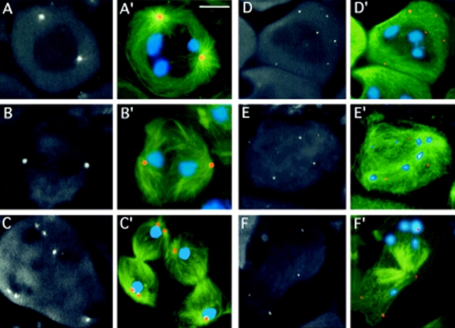
Failure of centrosome assembly in asl mutants. Cells were stained for α tubulin (green), γ tubulin (orange), and DNA (blue). Panels in black and white show only γ tubulin immunofluorescence; color panels show merged images. (A–C′) wild-type; (A, A′) prometaphase I; (B, B′) early anaphase I; and (C, C′) telophase I showing well-organized centrosomes that accumulate γ tubulin. In one of the telophases shown in C and C′, centrosomes have already started to separate in preparation for the second meiotic division. (D– F′) asl mutants; (D, D′) prometaphase I–like figure; (E, E′) anaphase I; and (F, F′) telophase I, showing no γ tubulin accumulations at the cell poles. Note that γ tubulin is dispersed in small aggregates that do not appear to have the ability to nucleate microtubules. Bar, 10 μm.

