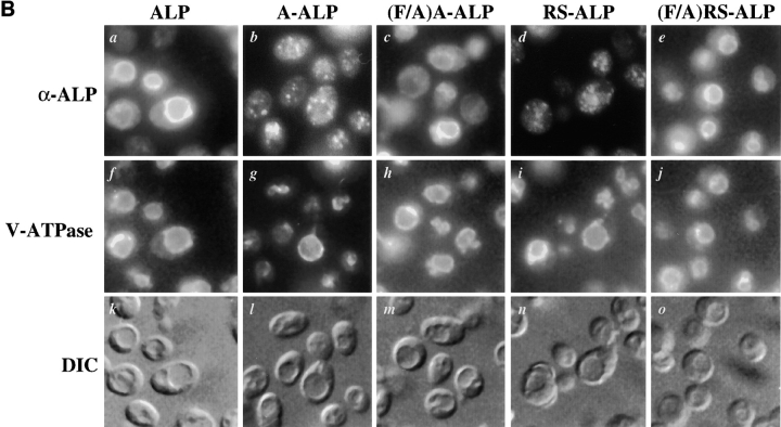
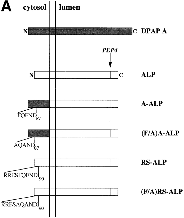
Figure 1.
(A) Schematic representation of proteins used in this study. Sequences derived from DPAP A are shown shaded and those from ALP are unshaded. (B) Localization of ALP, A-ALP, (F/A)A-ALP, RS-ALP, and (F/A)RS-ALP in wild-type cells. NBY72 (pho8Δ-X pep4-3) cells harboring pSN92 (ALP; a, f, and k), pSN55 (A-ALP; b, g, and l), pSN100 ((F/A)A-ALP; c, h, and m), pSN97 (RS-ALP; d, i, and n), or pSN123 ((F/A)RS-ALP; e, j, and o) were prepared for double labeling indirect immunofluorescence using the α-ALP mAb, 1D3-A10 (a–e) and affinity-purified antibodies against the 100-kD subunit of the V-ATPase, Vph1p, to show the localization pattern of the V-ATPase (f–j) as described in Materials and Methods. Cells were also visualized using DIC microscopy (k–o).


