Figure 2.

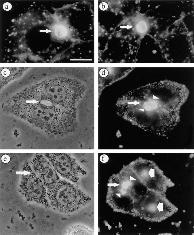
BC-derived C6-NBD-GlcCer and C6-NBD-SM accumulate in SAC at 18°C. Cells were labeled with 4 μM C6-NBD-GlcCer or C6-NBD-SM at 37°C for 30 min. Subsequently, the fluorescent lipid pool in the basolateral PM was removed by back exchange (a, C6-NBD-SM; b, C6-NBD-GlcCer; arrows point to BC). Alternatively, the cells were subsequently washed and further incubated at 18°C for 60 min. C6-NBD-SM (c and d) and C6-NBD-GlcCer (e and f) labeled the BCP of the cells, i.e., BC (thin arrows) and vesicular structures located near the BC membranes (arrowheads). In cells that did not participate in the formation of BC some accumulation of the fluorescent lipid analogue in juxtanuclear compartments was often seen (f, wide arrows) (c and e, phase contrast to d and f, respectively). Bar, 10 μm.
