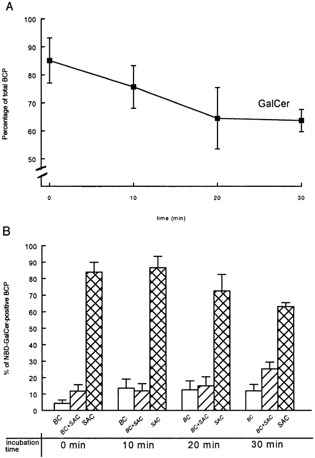
Figure 6.
Semi-quantitative kinetic analysis of C6-NBD-GalCer, exiting from SAC. The analogue was accumulated in SAC as follows. Cells were labeled with 4 μM C6-NBD-GalCer at 37°C for 30 min to label BC. After depletion of fluorescent lipids from the basolateral PM, the cells were subsequently incubated in HBSS + BSA at 18°C for 60 min. Cells were then treated with 30 mM sodiumdithionite at 4°C for 7 min to eliminate BC-associated fluorescence. After removal of the sodiumdithionite by extensive washing, the temperature was shifted to 37°C to reactivate transport from SAC. In A, the percentage of BCP labeled with C6-NBD-GalCer after a 0, 10, 20 and 30 min chase is presented. In B, the distribution of the BCP-associated C6-NBD-GalCer is shown. In A and B, data are expressed as percentage of total (i.e., NBD-positive + NBD-negative) BCP (mean ± SEM) and of total NBD-positive BCP of at least four independent experiments carried out in duplicate, respectively.

