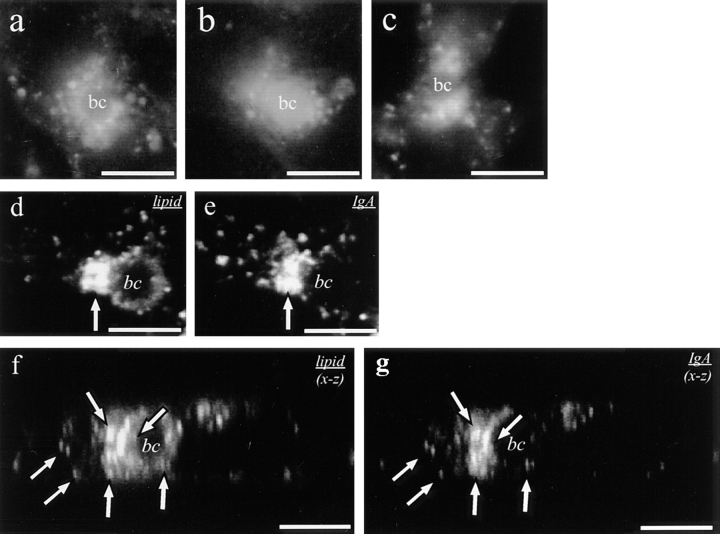
Figure 8.
Transcytosing IgA and NBD-labeled sphingolipids colocalize in SAC in HepG2 cells. Asialofetuin-pretreated cells were labeled with 50 μg/ml TxR-IgA at 4°C for 30 min, washed, and then incubated at 37°C for 20 min in HBSS (a). The cells were further incubated at 37°C for another 30 min (b) or alternatively, cooled to 18°C and further incubated at this temperature for 60 min (c). In d–g, confocal images of the BC area, labeled with C6-NBD-lipid (d and f) or TxR-IgA (e and g) are shown (f and g, x-z sections of d and e, respectively). Note the similarity in the shape of the fluorescence pattern obtained for accumulating protein and lipid, adjacent to the BC (arrows). Bar, 5 μm.

