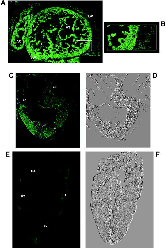
Figure 7.

Confocal image of the hearts at different stages of development. (A and B) Cross-sections of the heart from 14.5-d-old embryo. GFP expression is localized to both atrial and ventricular walls but the fluorescent signal is also seen in the atrial and ventricular trabeculae. (B) The highest fluorescent signal found in the cardiomyocytes is shown. (C) GFP expression in the heart of 18-d-old embryo. (D) A phase-contrast image of C. (E) A negligible level of expression of GFP in the heart of 3-wk-old mouse is shown. (F) A phase-contrast image of E. Symbols: AC, atrial chamber; B, blood cells; TW, thoracic wall; VT, ventricular trabeculae; and VW, ventricular wall.
