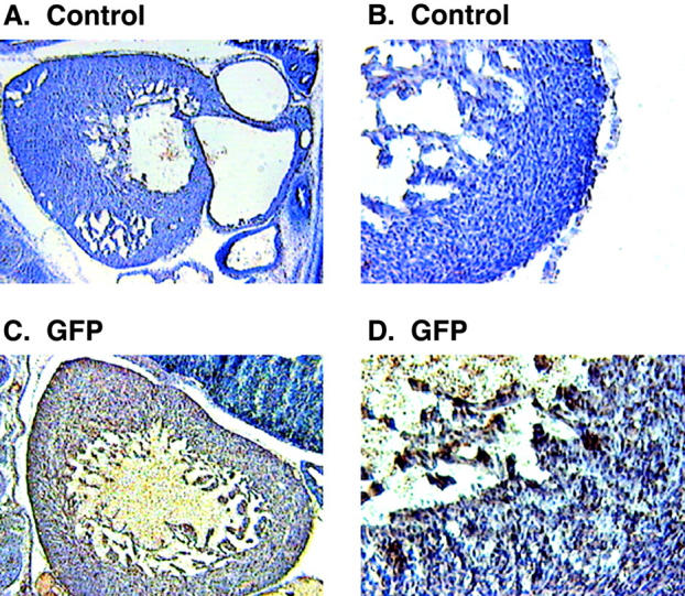
Figure 8.

Immunohistochemical analysis of the GFP transgenic mice. Sagittal sections of 13.5-d-old mouse embryos were stained with specific antibodies followed by DAB (brown color). The sections were counter-stained with hematoxylin to visualize the nuclei. Low (A) and high magnification (B) images from cryostat sections of a wild-type mouse embryo stained with anti-GFP antibody. Low (C) and high (D) magnification images from GFP transgenic mouse showing the GFP positive staining in the ventricular myocytes.
