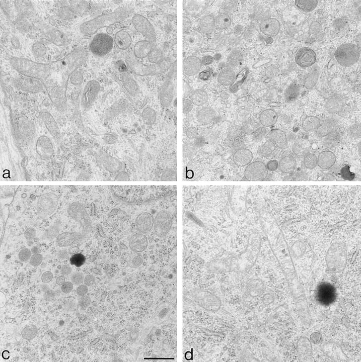
Figure 4.

Ultrastructural analysis of mitochondria by electron microscopy. Sympathetic neurons were cultured for 5 d in the presence of NGF. At day 5, some cultures were maintained in the presence of NGF for an additional 2 d (a), or deprived of NGF for 1 d (b) or deprived of NGF but in the presence of BAF (c). After 2 d of treatment with BAF, cultures of neurons protected from apoptosis were re-exposed to NGF for an additional 2 d (d). Bar, 0.5 μm.
