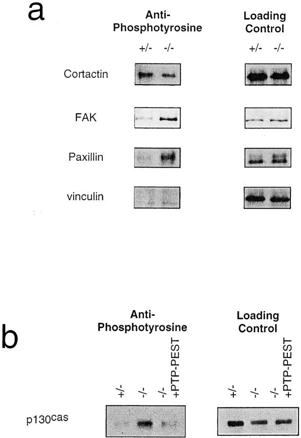
Figure 4.
(a) Constitutive hyperphosphorylation of FAK and paxillin in PTP-PEST (−/−) cells. Left column shows extent of tyrosine phosphorylation of immunoprecipitates as detected by the 4G10 antiphosphotyrosine mAb; right column shows evaluation of the amount of protein loaded using the same antibody as the immunoprecipitation. (b) Rescue of p130CAS hyperphosphorylation in PTP-PEST (−/−) overexpressing wild-type PTP-PEST. p130CAS was already shown to be a substrate for PTP-PEST (Garton et al., 1996) and to be hyperphosphorylated in the PEST (−/−) cells (Côté et al., 1998). Left panel shows phosphorylation levels of p130CAS in each cell line; right panel shows loading control.

