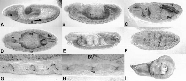
Figure 5.
Wb protein pattern during embryonic and larval development. Embryos are oriented with the anterior to the left and dorsal side up unless otherwise noted. (A) Stage 10 embryo (embryonic stages are those of Campos-Ortega and Hartenstein, 1985) denoting low level staining in the layer between ectoderm (e) and mesoderm (m; arrow), the anterior midgut (am), and the proctodeum (pr). (B) Stage 13 embryo showing Wb localized on visceral mesoderm of the gut. (C) Stage 14 embryo, BM surrounding the major organs of the presumptive digestive systems, pharynx (p), esophagus (e), midgut (mg), and hindgut (hg) show marked staining. (D) Same stage 14 as in C, horizontal section, muscle attachment (ma) sites are stained. (E) Stage 15 embryo, major BMs are stained. (F) Stage 16 embryo, horizontal section, BMs of the major portions of the digestive system show staining. (G and H) High magnifications of muscle attachment sites of stage 12 and stage 16 embryos, respectively. Note also the distinct staining of the BM of the midgut. (I) Wing disc, staining is particularly strong in distinct regions on the presumptive wing dorsal and ventral region.

