Figure 7.
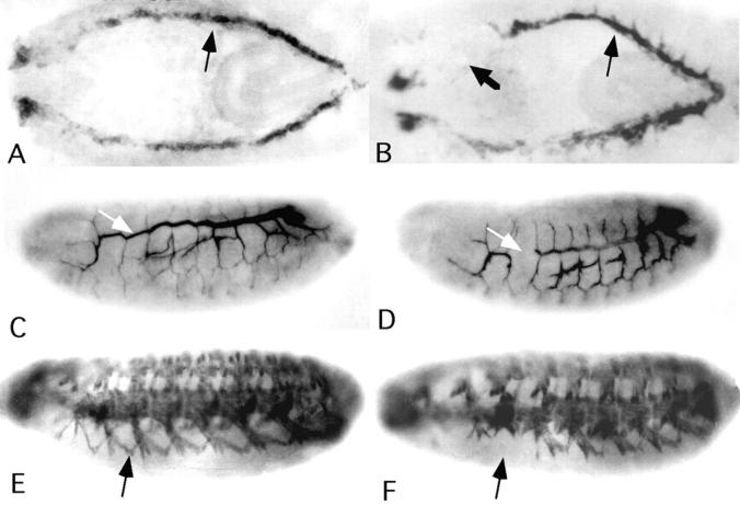
Dorsal vessel, trachea, and somatic muscle defects in wb embryos. Wild-type embryos (A, C, and E) and abnormal embryos from wbk05612 producing crosses (B, D, and F) are shown with anterior at the left. (A and B) Antibody staining of stage 14 embryos shows mislocalization (arrow) and gaps (thick arrow) in the pericardial cells of the developing heart and dorsal vessel of a wb embryo. (C and D) Antibody staining of stage 16 embryos shows gaps (arrow) in the dorsal tracheal trunk of a wb embryo in D. (E and F) Ventrolateral views of stage 16 embryos stained with antimyosin antibodies to visualize somatic muscle patterning. Ventral oblique muscles (arrow) in the wb embryo of F never reach their targeted epidermal attachment sites.

