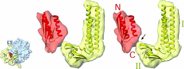
Fig. 4.
Stereoview presentation of the interaction between CTD of PSRP1 with domain II of pRRF. Difference map, showing the N-terminal domain of PSRP1 (semitransparent red), is shown with the fitted homology model (red ribbons). N-terminal (N) and C-terminal (C) ends are labeled. Difference map corresponding to pRRF (semitransparent yellow) is shown with fitted atomic structure (greenish-yellow ribbons) of Thermotoga maritima RRF (PDB ID 1DD5). Two domains (I and II) of RRF are labeled. Thumbnail to the left depicts the orientation of the chloro-ribosome with small (semitransparent pale yellow) and large (semitransparent blue) subunits identified.

