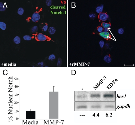
Fig. 7.
MMP-7 activity leads to nuclear translocation of N1ICD and expression of hes-1. (A and B) COS-7 cells expressing full-length Notch-1 with a C-terminal V5 tag were treated for 4 h with MMP-7. Shown is immunofluorescence for Cleaved N1 Val-1744 antibody (green) and V5 antibody (red). (Scale bar, 20 μm.) Cells were treated for 4 h with medium alone (A) or with rMMP-7 (B). Arrows indicate cells with nuclear cleaved Notch-1. DAPI is shown in blue. (C) Quantification of Notch-1 nuclear translocation. Percentage represents cells with nuclear Notch-1 divided by total cells expressing Notch-1. (D) RT-PCR for hes1 from COS-7 cells expressing Notch-1 treated with MMP-7 or EDTA. Numbers represent fold expression relative to medium alone. Data are representative of five independent experiments.

