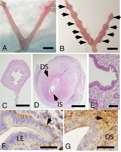
Fig. 3.
PEGLA blocks blastocyst implantation and immunolocalizes to uterine luminal epithelium on D3.5. Shown are representative photomicrographs of uteri collected on D6.5 from mated mice treated with 12.5 mg/kg (25 μl) i.p. PEGLA (A) or control (mPEG2-NHS) (B) at 1200 hours and 2200 hours on D2.5 and 1000 hours on D3.5. Arrows indicate implantation sites. (C) H&E-stained cross section of A. (D) H&E-stained cross section of implantation site in B. (E) Higher magnification of C showing normal (intact) epithelial and stromal architecture after PEGLA administration. (F and G) PEG immunostaining in uteri of two different mice on D3.5 after i.p. administration of 12.5 mg/kg (25 μl) PEGLA at 1200 hours and 2200 hours on D2.5 and 1000 hours on D3.5. Arrows indicate positive staining as described in Results. (DS, decidualized stroma; IS, implantation site; LE, luminal epithelium. (Scale bars: A and B, 5 mm; C and D, 0.5 mm; E–G, 50 μm.)

