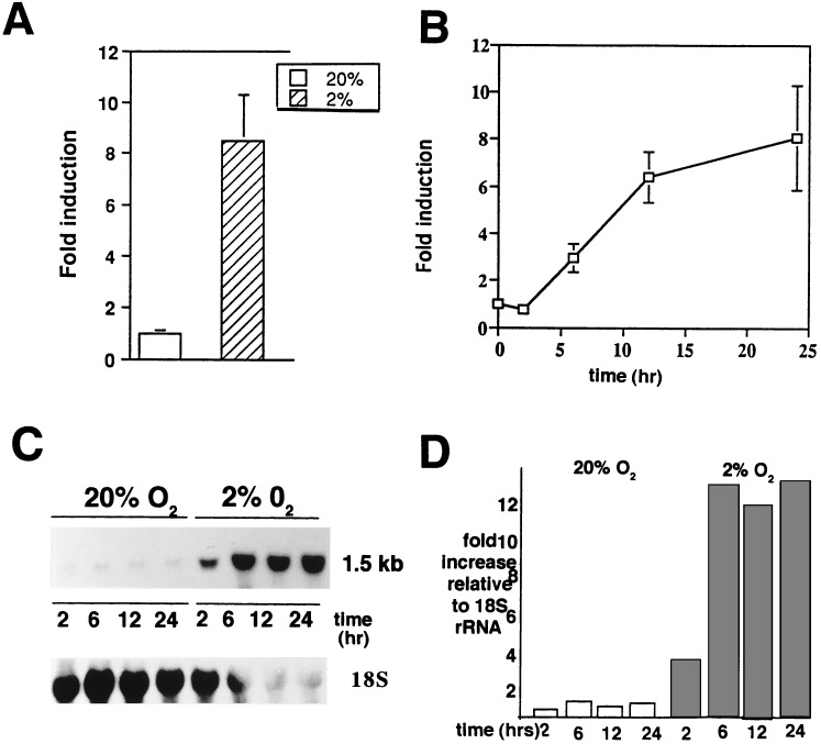
Figure 1.
Hypoxia induction of IGFBP-1 in HepG2 cells. (A) Cells were grown in serum-containing medium in the presence of 20% and 2% O2, respectively. After 24 hr, the conditioned medium was collected and assayed in triplicate for IGFBP-1 using an immunoradiometric assay. Values shown are the mean ± SEM of six different experiments. (B) Time course of hypoxia experiments, with media being harvested after 2, 6, 12, and 24 hr of exposure of HepG2 cells at 2% and 20% pO2. Data represent the mean of four experiments run in triplicate and are reported as “fold induction.” (C) At the end of each time period, total RNA was collected from the cells and a 1.5-kb mRNA transcript was detected by Northern blot analysis using an EcoRI fragment (938 bp) of the IGFBP-1 cDNA. 18S rRNA shown at the bottom. (D) Densitometry of Northern blot shown in C, with fold increase normalized to 18S rRNA.

