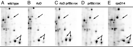
Fig. 2.
Two-dimensional electrophoresis of total cell extracts from wild-type MAS70 (A), ME97 rluD::cat dust (B), ME123 rluD::cat dust, prfBE172K (C), ME125 rluD+prfBE172K (D), and the ram strain rpsD14 (E) (31). Sections of the gels showing protein with size from 55,000 to 30,000 (top to bottom) and isoelectric points from 6 to 4.5 (left to right). The position of a spot with the molecular weight and isoelectric point of RF2 is indicated with an arrow. The double arrow points to the outer membrane proteins OmpC and OmpF. Especially in this area mistranslation in the ram strain can be seen (E).

