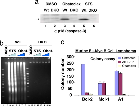
Fig. 2.
Induction of apoptosis and inhibition of colony formation by obatoclax. (a) Deletion of Bax and Bak causes resistance to obatoclax. Baby kidney epithelial cell lines derived from the WT and Bax,Bak double knockout (DKO) mouse were treated with vehicle (DMSO) or vehicle containing 2.0 μM obatoclax or 0.5 μM staurosporin (STS), and 20 h later the processed large subunit of caspase-3 (arrow) was detected by SDS/PAGE and immunoblot analysis. (b) As in a, except that oligonucleosomal fragments were detected by agarose gel electrophoresis and staining with ethidium bromide after treatments with 0.05 μM (lanes 3 and 10), 0.2 μM (lanes 4 and 11) and 1 μM (lanes 5 and 12) STS for 16 h; obatoclax treatment was 0.5 μM (lanes 6 and 14), 1 μM (lanes 7 and 15) and 2 μM (lanes 8 and 16) for 24 h. (c) Sensitivity of murine Eμ Myc B cell lymphoma cell lines overexpressing Bcl-2, Mcl-1, or A1 to obatoclax. Cells were exposed to 1 μM obatoclax or ABT737 for 24 h, the media (containing compound) diluted 30-fold, and colony formation on soft agar recorded after 7 days, as described in ref. 45.

