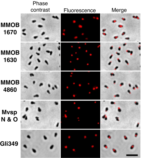
Fig. 6.
Subcellular localization of protein components of the jellyfish structure examined by immunofluorescence microscopy. The labeled proteins are indicated at the left by their gene IDs. Phase contrast, fluorescence and merged images are presented in Left, Center, and Right, respectively. The cells were chemically fixed, permeabilized by 0.1% Triton X-100, and labeled for the proteins other than Gli349. The permeabilizing step was not applied for labeling of Gli349. MMOB1670, MMOB1630, and MMOB4860 proteins correspond to the bands b, f, and j, respectively, shown in Fig. 4. The amino acid sequence of MvspN shared 314 aa with the MvspO sequence of 458 aa, and the antibody used here is known to recognize both proteins equally in Western blotting (17). (Scale bar, 2 μm.)

