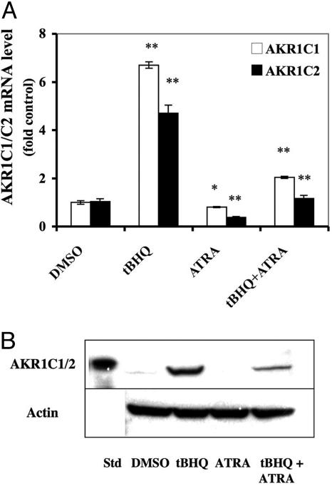
Fig. 2.
Inhibition of AKR1C1/2 induction by ATRA in AREc32 cells. AREc32 cells were incubated with DMEM containing either DMSO, tBHQ (10 μM), ATRA (1 μM), or tBHQ (10 μM) plus ATRA (1 μM) for 24 h. (A) AKR1C1 and C2 mRNA analysis. AKR1C1 and AKR1C2 mRNA were measured by TaqMan analysis; the level of 18S rRNA was used as an internal standard. Control cells were treated with DMSO only. The TaqMan data show mean ± SD from triplicate samples and represent the results of two separate experiments. *, P < 0.05; **, P < 0.005. (B) Repression of tBHQ-mediated induction of AKR1C1/2 protein expression by ATRA. AREc32 cell extracts were prepared and the expression of AKR1C1/2 was measured by Western blotting. The experiment was repeated three times with similar results.

