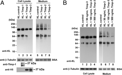
Fig. 3.
Effects of Timp-1, Timp-2, and Timp-3 on KL shedding. (A) COS-7 cells were transfected with KL alone (KL control) or cotransfected with KL and Timp1, Timp-2, or Timp-3 as indicated. Forty-eight hours after transfection, the cells were washed and incubated in serum-free medium as described before. The protein samples in the cell lysates and medium were collected and analyzed by Western blotting. BSA and tubulin were used as loading controls. The expression of Timp-1 was analyzed by Western blotting with anti-Timp-1 antibody, and the expressions of Timp-2 and Timp-3 were analyzed with an anti-V5 antibody. The apparent molecular masses of the bands are indicated. Statistical analysis of the densitometric results is shown in SI Text. (B) Forty-eight hours after transfection, KL-transfected COS-7 cells were incubated in serum-free DMEM without (KL control) or with various amounts of the Timp-3 peptide, as indicated. The proteins in the cell lysates and medium were analyzed as described in Fig. 1.

