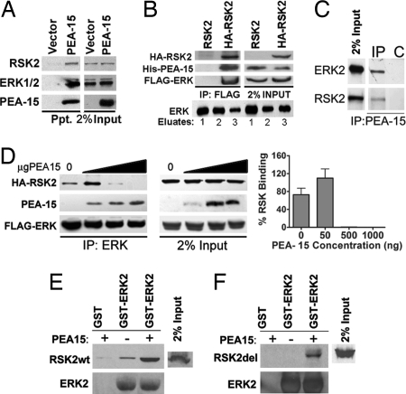
Fig. 1.
PEA-15 enhances ERK1/2 association with RSK2. (A) HeLa cells were transfected with His6-tagged PEA-15 or empty vector. Cell lysates were passed over a Probond Ni2+ column. Protein precipitates were immunoblotted to detect endogenous RSK2, ERK1/2, and His-PEA-15. (B) (Upper) Cos-7 cells were transfected with FLAG-ERK2, His-PEA-15, and HA-RSK2 or untagged RSK2 control. (Lower) Lysates were pooled and precipitated with FLAG-ERK2. FLAG-ERK was eluted off the column with FLAG peptide. HA-RSK2 was immunoprecipitated from the eluate. The immune complexes were immunoblotted for PEA-15, FLAG-ERK2, and HA-RSK2. (C) Endogenous PEA-15 was immunoprecipitated (IP) from primary astrocytes. Beads alone (C) were used as control. The precipitate was immunoblotted for ERK and RSK2. (D) Cos-7 cells were transfected with equal amounts of HA-RSK2 (1 μg) and FLAG-ERK2 (1 μg) and increasing amounts of His-PEA-15 plasmid (0, 50, 500, or 1,000 ng). FLAG-ERK2 was immunoprecipitated, and the precipitated complexes were immunoblotted to identify RSK2, ERK1/2, and PEA-15. Control immunoblots indicate 2% of the input protein levels. The graph of the percentage of RSK2 precipitate was derived from spot densitometry of three independent experiments. (E and F) Cos-7 cells were transfected with RSK2 (RSK2wt) or a deletion mutant of RSK2 (RSK2del) that does not bind ERK. Lysates of these cells were run over GST-ERK in the presence or absence of 300 μg of PEA-15 protein. Bound RSK2 was determined by immunoblot with anti-HA antibody. (E) Western blots showing full-length RSK2 bound to ERK with and without PEA-15. (F) Western blot showing the amount of RSK2 deletion mutant (Rsk2del) bound to GST-ERK2.

