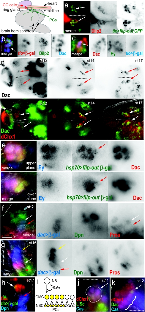
Fig. 1.
IPC lineage development in the Drosophila brain. The diagram, at top left, shows the positions of the IPCs, within in the pars intercerebralis, and the CC cells within the ring gland, relative to the embryonic and larval protocerebrum. All brains labeled by antibodies are as indicated with the text color corresponding to color channels in merged images. The position of the midline is indicated by dotted lines with anterior to the left when midline is horizontal, or anterior to the top when midline is vertical. Embryonic stages of labeled brains are as indicated. For an indication of scale, note that the individual cells of the IPC lineage are typically 3–8 μm at all stages. (a) Dorsal view of IPCs in third-instar larval brain shows the specificity of the tio enhancer for IPCs (arrows). (b) IPCs in the pharate larval brain express tio and Dac (arrow). (c) Before onset of Dilp2 expression, tio+ cells comprise the anterior part of a 10- to 12-cell cluster of Dac+ Ey+ cells (arrow). (d) The expansion of the Dac+ IPC lineage from one cell at stage 12 to 10–12 cells by stage 17 (arrows). (Upper) Series shows Dac expression only. (Lower) Series shows the Dac+ cluster expands on the posterior-lateral side of the anterior PI (6), marked by dChx1. (e) The Dac+ IPC lineage is marked by a single marked clone at early stage 17 (red arrow). The two focal planes show the entire Dac+ group. Ey expression appears in the older anterior cells of the Dac+ group at this stage. (f and g) The IPC lineage is produced from a dividing Dpn+ Dac+ neuroblast with membrane-localized Pros (red arrow). At stage 12 (f), neuroblast has divided once and the GMC daughter cell shows nuclear localized Pros (white arrow). At stage 16 (g), the neuroblast is still present (red arrow), as are the GMCs (white arrow). Cells to the anterior of the Dac+ group are early IPCs, lacking both Dpn and Pros expression (yellow arrow). (h) IPCs arise from the first-born GMCs. tio expression (white arrow), which labels the IPCs, is in the anterior of the Dac+ group, whereas the IPC neuroblast is at the posterior (red arrow). (i) Lineage model for IPCs. (j) The dChx1+ Cas+ L'Sc+ proneural group, which gives rise to the IPC neuroblast, in the anterior neuroectoderm (area in dotted outline, arrow). (k) The dChx1+ Cas+ cells delaminate as a chain of neuroblasts (bracket). The first to delaminate is the Dac+ IPC neuroblast (arrow). (Scale bar: 5 μm.)

