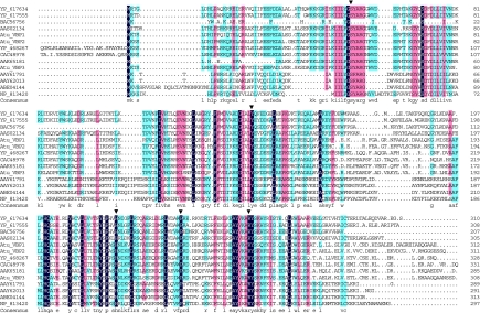
Fig. 1.
Sequence alignment of VBP homologs. The VBP homologs and the bacterial species encoding them are described in SI Table 1. The identical and conserved residues are boxed in black and colors, respectively. The arrowheads indicate the VBP1 point mutations at amino acid positions 52 (S52A), 139 (Y139A), 218 (H218A), 239 (P239A), and 260 (R260A). Atu, A. tumefaciens.

