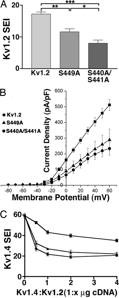
Fig. 4.
Mutations at in vivo phosphorylation sites suppress Kv1.2 surface expression and Kv1.2 currents. (A) COS-1 cells transfected with WT Kv1.2, S440A/S441A, or S449A were double immunofluorescence stained with Kv1.2e before and K14/16 after permeabilization, and a SEI determined the percentage of Kv1.2-expressing (K14/16-positive) cells with Kv1.2e surface staining. Statistical significance was determined by one-way ANOVA followed by Turkey's post hoc test, and statistical significance was considered at *, P < 0.05; **, P < 0.01; and ***, P < 0.001. (B) Whole-cell patch–clamp recordings from HEK293 cells expressing WT Kv1.2 (squares), S440A/S441A (circles), or S449A (triangles). The cells were held at −80 mV and step depolarized to +60 mV for 200 ms in +10-mV increments. Peak current amplitudes at each test potential were divided by the cell capacitance to obtain the current densities. Mean ± SE of current densities obtained (Kv1.2, n = 11; Kv1.2 S440A/S441A, n = 6; S449A, n = 4) were plotted against each test potential. (C) Dose-dependent effects on Kv1.4 surface expression in the presence of increasing amounts of WT Kv1.2, S440A/S441A, or S449A cDNA in COS-1 cells (n = three samples of 100 cells each). Symbols are the same as in B.

