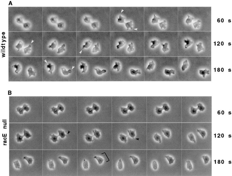
Figure 1.

Dictyostelium wild-type and racE null cells complete cytokinesis normally when attached to a substrate. Dictyostelium wild-type (strain DH1) and racE null (strain 24EH6) cells were allowed to attach to coverslips and were visualized by video light microscopy. (A) Wild-type cells form a cleavage furrow that divides the cells 3–5 min after furrow formation. As the daughter cells separate, they form large pseudopods (white arrowheads) to migrate away from each other. (B) RacE null cells also form a cleavage furrow that successfully divides the cell. However, the daughter cells do not form large pseudopods. Instead, they have multiple filopodia (white arrowheads) and a broad leading edge (bracket). The time between each frame is 10 s. QuickTime movies of the images in this manuscript can be accessed at http://note.cellbio. duke.edu/Faculty/∼http://DeLozanne/Gerald1998a.html
