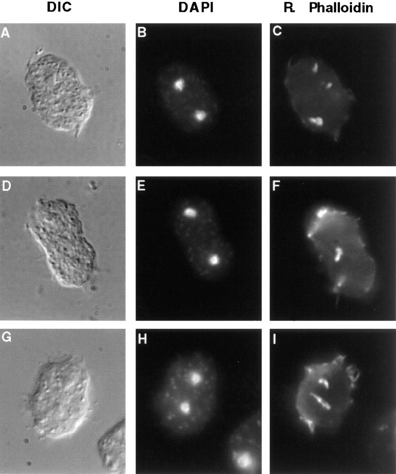
Figure 10.

RacE null cells have abnormal F-actin inclusions. Cells were fixed in suspension and stained as in Fig. 9. These three cells are examples of about 40% (n = 42) of mitotic racE null cells that display abnormal F-actin aggregates. These inclusions were not observed in racE null cells during interphase or in any wild-type or myosin II null cells at any stage of the cell cycle. The inclusions appear to form only as racE null cells attempt to divide in suspension.
