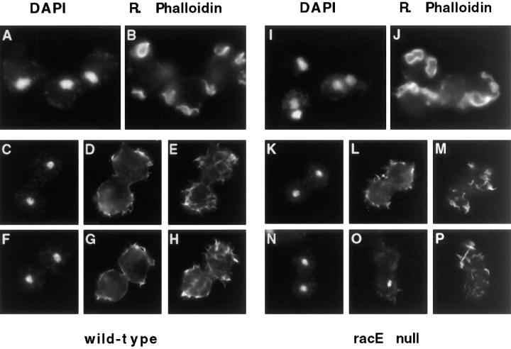
Figure 9.
F-Actin is reorganized in mitotic Dictyostelium cells but is not concentrated in the cleavage furrow. Cells were fixed in suspension cultures and stained with DAPI (A, C, F, I, K, and L) and rhodamine–phalloidin (B, D, E, G, H, J, L, M, O, and P). During interphase (recognized by the large, decondensed nuclei) F-actin is concentrated in filopodia, crowns, and ruffles of both wild-type and racE null cells (A and B; I and J). During mitosis, both cell types loose their crowns and ruffles and are covered by many filopodia (C–H and K–P). F-actin is not particularly concentrated at the cleavage furrow of dividing cells. Two focal planes are shown for mitotic cells stained with rhodamine phalloidin. (A–H) Wild-type cells; (I and J) racE null cells.

