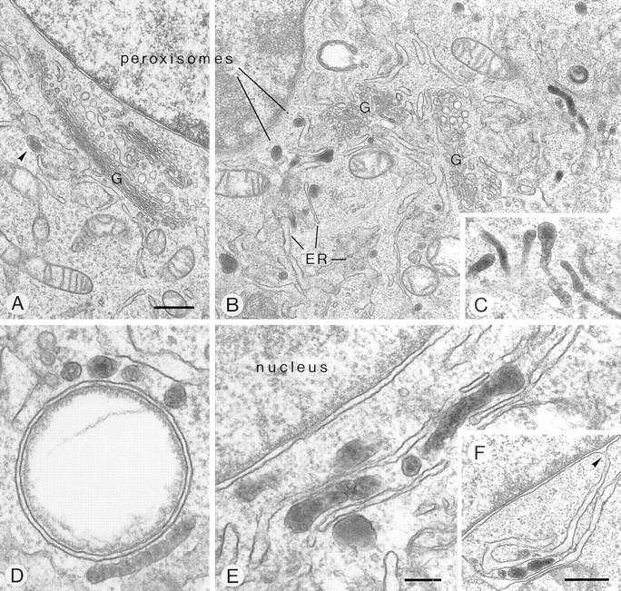
Figure 10.

Fine structure of peroxisomes in the perinuclear region of mutant ldlF cells at permissive (34°C, A, arrowhead) and nonpermissive (39.5°C, B–F) temperature. Phenotypic peroxisome clustering and formation of peroxisome-ER aggregates is revealed, due to functional deficiency of ε-COP. At nonpermissive temperature, the intensely DAB-stained peroxisomes frequently exhibit a highly tortuous, tubular shape (B–E). They cluster at the periphery of lipid vacuoles (D) and form rows alternating with ER cisternae (E), the latter of which are often in continuity with the perinuclear membrane (F, arrowhead). G, Golgi complex. Bars: (A–C and F) 500 nm; (D and E) 150 nm.
