Figure 9.
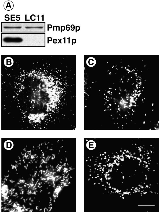
Phenotypic change in peroxisomal morphology by overexpression of Pex11p (A–C) as well as functional deficiency of coatomer (D, E). CHO cells were stably transfected with either Pex11p-cDNA cloned into pcDNA3 (SE5) or pcDNA3 lacking an insert (LC11). Carbonate membranes of postnuclear supernates (10 μg) were analyzed for their content of Pmp69p and Pex11p by SDS-PAGE and Western blotting (A). The peroxisomal compartment of SE5 (B) and LC11 (C) was visualized by immunofluorescence using the polyclonal anti-Pmp69p antiserum. ldlF cells expressing a ts mutant of ε-COP were kept for 24 h at nonpermissive (39.5°C, D) or permissive (34°C, E) temperature before immunofluorescence staining of peroxisomes using the anti-Pmp69p antiserum. Note numerical increase in small spherical peroxisomes (B) and clustering of tubular peroxisomes (D). Bar, 3 μm.

