
Fig 2.
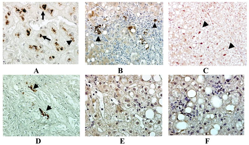
M-30 was present in MDBs (arrows) in NASH patients’ liver biopsies (A). M-30 stained aggresomes were present in hepatocytes and macrophages (arrow heads) (A-D). Some livers were negative for any positive staining for M30 in the presence of steatosis (E) or steatohepatitis (F). Immunoperoxidase ×260 (B, C, D), ×520 (A, E, F).