
Fig. 4.
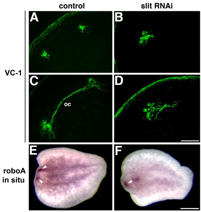
Midline defects appear at early stages of regeneration after Smed-slit RNAi. (A-D) Confocal projections of VC-1 immunofluorescence showing regeneration of the photosensitive cells in control (A,C) and Smed-slit RNAi animals (B,D). (A) In control planarians, two bilateral clusters of photosensitive cells are detected between days 2 and 3 of regeneration; this image corresponds to a 3-day blastema. (C) Between days 3 and 4, axons project contralaterally to form the optic chiasm (oc); this image corresponds to a 4-day blastema. (B) After Smed-slit RNAi a single cluster of photosensitive cells is observed within the blastema. (D) In Smed-slit RNAi animals axons project from the photosensitive cells, forming a greatly reduced chiasm at the midline. (B) and (D) show 3-day blastemas. (E-F) Regenerating cephalic ganglia visualized by whole-mount in situ hybridization to detect Smed-roboA (Cebrià and Newmark, 2007) in regenerating tail pieces 2 days after amputation. (E) Two bilateral clusters of cells (arrowheads) constitute the brain primordia that develop within the regeneration blastema between days 1 and 2. (F) The brain primordia collapse at the midline after Smed-slit RNAi: a single cluster (arrowhead) of new brain cells is observed within the blastema. (A-D) anterior to the top left corner. (E-F) anterior to the left. Scale bars: 50 μm in A-D; 250 μm in E, F.