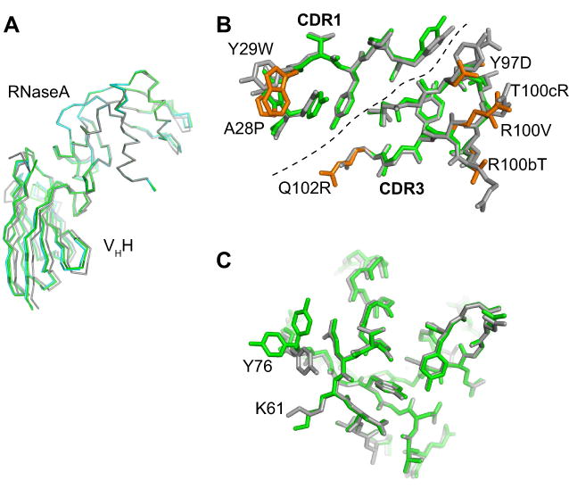
Figure 3. High-resolution x-ray crystal structures of wild-type and affinity-matured VHHs in complex with RNaseA.

(A) A comparison of the two structures after superposition of the RNaseA portion. The wild type is shown in gray and the mutant in green. (B) CDR1 and CDR3 residues of the wild-type (gray) and mutant (green) VHHs. The dashed line divides CDR1 and CDR3. Mutations in the affinity-matured VHH are shown in orange and labeled. (C) The epitope on RNaseA in the wild-type (gray) and mutant (green) complexes. Y76 has two conformers in the mutant structure.
