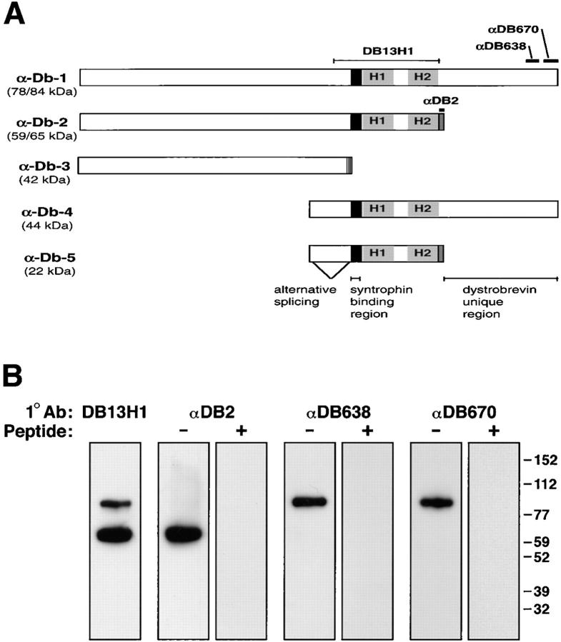
Figure 1.
Antibodies to α-dystrobrevin isoforms in skeletal muscle. (A) Schematic of the known α-dystrobrevin cDNAs identified in mouse (10) and human tissues (35). The coding region includes a syntrophin-binding region (for review see reference 20) and a pair of coiled-coils (9). Abs were generated against peptides (shown as bars) corresponding to regions specific for isoforms. Predicted molecular weights for mouse α-dystrobrevin-1 and -2 were calculated from the mouse sequences while α-dystrobrevin-3, -4, and -5 were estimated based on the corresponding sequences in human. (B) The specificities of dystrobrevin antibodies were tested by immunoblot analysis. Dystrobrevin–dystrophin–syntrophin complexes were partially purified with mAb SYN1351 from Triton X-100–solubilized skeletal muscle extracts and immunoblotted with affinity-purified antibodies prepared against sequences specific to α-dystrobrevin-2 (αDB2) or α-dystrobrevin-1 (αDB638, αDB670). Labeling with antibodies (−) was eliminated by preincubation of the antibody with antigenic peptide (+). Molecular weight markers are shown in kD.

