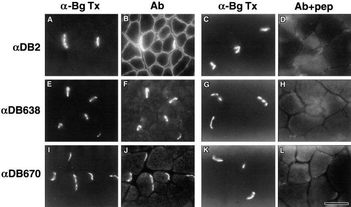
Figure 2.
Localization of α-dystrobrevin isoforms in skeletal muscle. The distribution of α-dystrobrevins in gastrocnemius skeletal muscle was compared with α-bungarotoxin (α-BgTx) staining of AChRs (A, C, E, G, I, and K). α-Dystrobrevin-2 antibodies (αDB2) stained the entire sarcolemma with particular concentration at the NMJ (B). Abs αDB638 and αDB670 directed against COOH-terminal tail of α-dystrobrevin-1 labeled NMJs with little or no extrasynaptic sarcolemmal labeling (F and J). Labeling for each antibody was dramatically reduced by preincubation of antibody with the appropriate antigenic peptide (D, H, and L). Bar, 50 μm.

