
Figure 6.
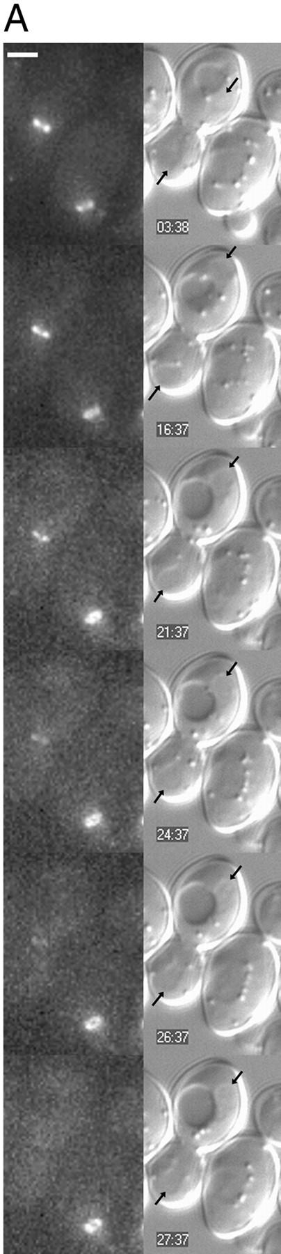
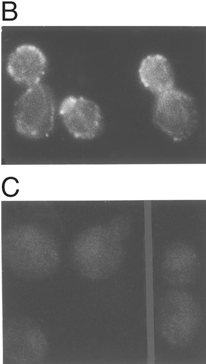
Dependence of Myo1p ring contraction, but not of Myo1p ring maintenance, on F-actin. (A) Cells of strain YEF1698 growing exponentially in SC medium were treated with 200 μM LAT-A for 10 min and then spotted onto solid SC/25% gelatin medium containing 200 μM LAT-A and observed by time-lapse video microscopy. Pairs of GFP fluorescence (left) and DIC (right) images were recorded at the times indicated. Arrows in DIC images, ends of the late-anaphase nucleus. Recording was continued for an additional 35 min beyond the images shown without obvious change in the appearance of the Myo1p–GFP ring in the right-hand cell. (B and C) Rhodamine-phalloidin staining of cells before (B) or 10 min after (C) the beginning of LAT-A treatment. Bar, 2 μm.