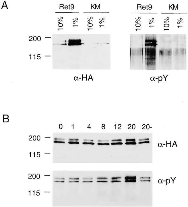
Figure 1.
(A) Autophosphorylation of RET in response to low serum. Ret9 and KM cells were cultured in 10 or 1% serum and then RET was immunoprecipitated with anti-HA and detected with anti-HA or anti-pTyr. (B) Ligand-dependent phosphorylation of RET. Ret9 cells cultured in 10% serum and 100 ng/ml GFRα-1 were exposed to 50 ng/ml GDNF for the indicated times in hours; the control culture 20− did not receive GFRα-1 or GDNF.

