
Figure 6.
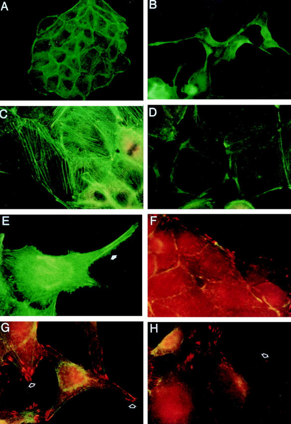
Effects of RET Activation on the actin cytoskeleton and focal adhesions. Ret9 cells cultured in media alone (A, C, and F) or with GDNF/GFRα-1 (B, D, E, G, and H). Cells were stained with FITC-phalloidin or antivinculin (red). Untreated cells exhibit prominent stress fibers (A and C), whereas RET activation results in fewer stress fibers and actin accumulation in lamellipodia (B) and at focal points along the cell membrane (D). Actin staining also reveals many filapodia (E, arrow) along the membrane of RET-activated cells. Control cells exhibit vinculin staining (F, red) at the termini of stress fibers. RET activation (G and H) results in vinculin staining at the leading edges of newly formed lamellipodia (arrows).