Figure 6.
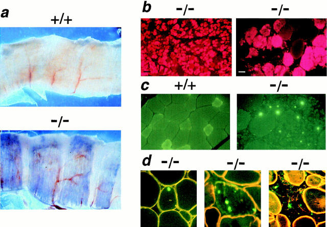
Vital staining with Evans blue dye (EBD) reveals disruption of membrane integrity in sarcoglycan-deficient muscle. Mice were injected with EBD and killed after 12 h. (a) Low-power magnification (5×) of diaphragm muscle from EBD-injected wild-type and mutant mice. Diaphragm muscle from gsg −/− mice exhibits segmental EBD uptake (seen as blue areas) while no EBD uptake is seen in wild-type diaphragm. (b) Regions of EBD uptake are seen as red cytoplasmic staining on fluorescence microscopy. No evidence of EBD microscopic staining is seen in gsg +/+ muscle (data not shown). (Left) a region of gsg −/− triceps muscle with marked EBD staining (black bar, 100 μm). (Right) A higher power view of gsg −/− gastrocnemius muscle showing EBD staining (white bar, 25 μm). (c) Apoptosis in γ-sarcoglycan–deficient muscle. TUNEL-labeling was performed on quadriceps muscle from a gsg −/− mouse. TUNEL-positive nuclei appear green. No TUNEL-positive nuclei were found in normal gsg +/+ myofibers (left). In contrast, myofibers with TUNEL-positive nuclei were seen commonly in gsg− /− muscle (right). (d) Counterstaining with an anti-dystrophin antibody (yellow-orange) demonstrates that TUNEL-positive nuclei are found within gsg− /− myofibers. Myofibers undergoing degeneration and demonstrating decreased membrane staining for dystrophin also show evidence of apoptosis.

