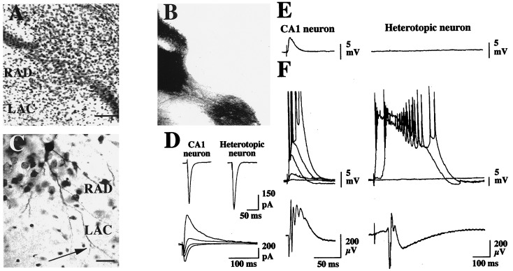
Figure 3.
Neocortical heterotopias receive hippocampal inputs. (A) Cytoarchitectonic staining of an heterotopia in a MAM brain in which a crystal of 1,1′-dioctadecyl-3,3,3′,3′-tetramethylindocarbocyanine perchlorate was inserted in CA3. Star indicates the position of the cell depicted in C. Afferent fibbers from CA3 to CA1 that are found in RAD avoid the heterotopia (B) with only sparse fibbers coursing in the ventral part of the heterotopic core. (Bar = 50 μm.) (C) The dendrites of biocytin-filled heterotopic neurons that extend up to the stratum lacunosum are putative targets for CA3 fibbers avoiding the heterotopic core. (Bar = 20 μm.) (D) Electrical stimulation of the Schaffer collaterals evoked an EPSC in both CA1 and heterotopic neurons (VM = −65 mV). When recorded at different holding potentials in heterotopic neurons, an EPSC/inhibitory post synaptic current sequence was observed in most cases (lower traces, from the most inward to the outward current, holding potentials were −70 mV, −60 mV, −50 mV, −40 mV). (E) Simultaneous whole cell patch clamp recordings of a pair of CA1 and heterotopic cells. Electrical stimulation of the Schaffer collaterals evoked an EPSP only in CA1 cell in control conditions (VM = −65 mV). (F) Same cells as in E. In the presence of bicuculline (10 μM), the same stimulation evoked a graded burst of action potentials in the CA1 cell and an all-or-none paroxysmal depolarizing shift in the heterotopic cell (upper traces). The corresponding field potentials are shown in the lower part. The shape of the evoked field potential was constant from slice to slice when the extracellular electrode was situated in the continuity of the CA1 pyramidal layer (see Fig. 4A).

