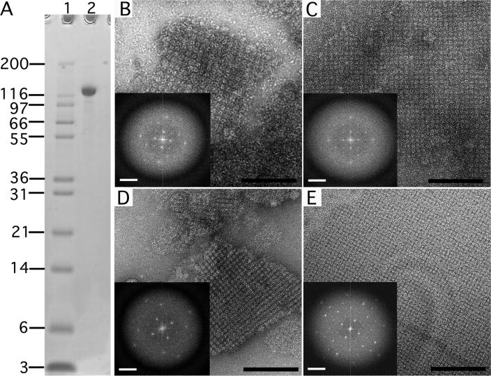
Figure 2. Negatively stained sbpA crystals formed in solution and on a lipid monolayer.
(A) SDS PAGE gradient gel showing purified sbpA used for crystallization. Lane 1: Mark 12 protein standard (Invitrogen Corporation, Carlsbad, CA); lane 2: sbpA (MW ∼120 kDa). (B) sbpA crystals formed in solution within 2 hours after addition of 50 mM CaCl2. (C) After incubation for 24 hours, the sbpA crystals grew larger but the order of the sbpA arrays did not improve significantly (compare Fourier transforms shown as insets in panels B and C). (D) sbpA crystals also formed within 2 hours of incubation with CaCl2 on lipid monolayers. (E) After a 24 hour incubation, the crystals grew not only bigger but also improved in order (compare Fourier transforms shown as insets in panels D and E). Scale bars in the images are 100 nm; scale bars in the Fourier transform are (6.5 nm)−1.

