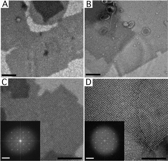
Figure 3. Negatively stained sbpA monolayer crystals prepared with the direct and loop transfer methods.
(A) Low magnification image (8,700×) and (C) high magnification image (52,000×) of sbpA monolayer crystals prepared with the direct transfer method. (B) Low magnification image (8,700×) and (D) high magnification image (52,000×) of sbpA monolayer crystals prepared using the loop transfer method. The insets in panels C and D show the Fourier transforms of the crystals. Scale bars in the images are 100 nm; scale bars in the Fourier transform are (6.5 nm)−1.

