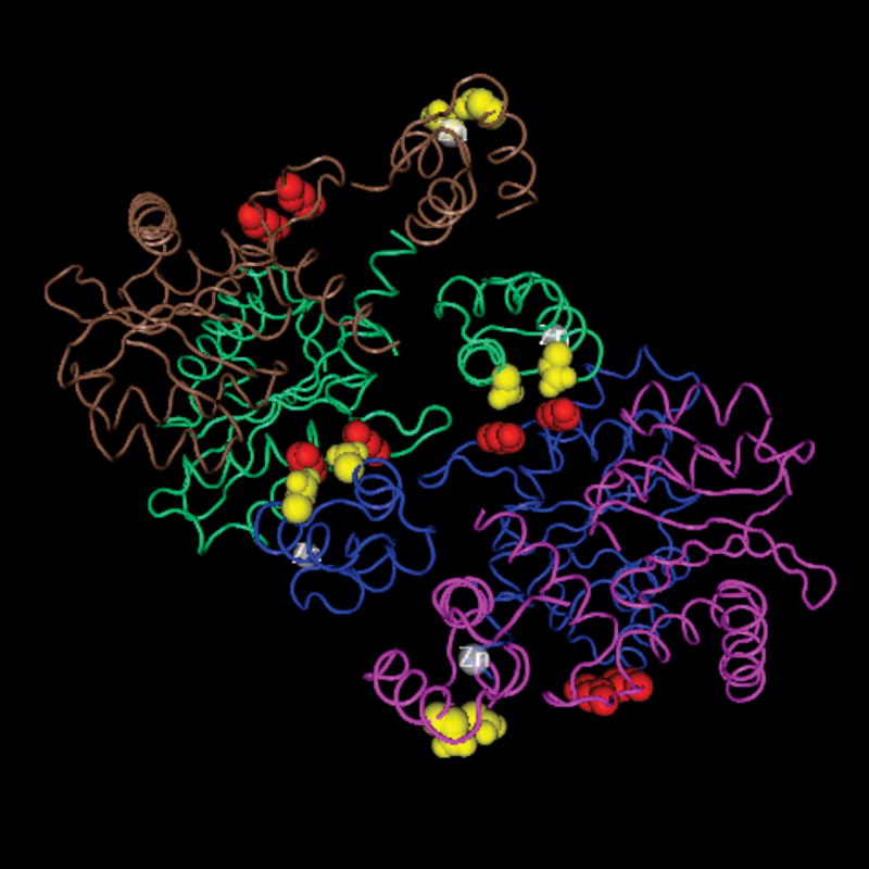
Fig. 1. KRK(186-188) and IN tetramerization.
The image was created in Cn3D, using the coordinates (accession number: 1K6Y) published by Wang and Craigie (Wang et al., 2001). Each of the four monomers of IN (N-terminal and core domains) is shown in a different color (brown, green, blue, purple), and the zinc ion is in white. For each monomer, the side chains of lysine 186 and lysine 188 are shown in red, while the side chains of glutamic acid 11 and aspartic acid 25 are shown in yellow.

