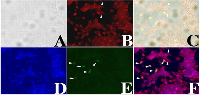
Figure 4.
Expressing PRPF31-GFP protein in primary retinal cells. Primary retinal cell cultures were established as described in Materials and Methods. A calcium-phosphate method was established to transfect primary retinal cells. Twelve hours after transfection, cells were fixed and immunostained using anti-rhodopsin antibody, and images were captured. A phase-contrast image (A), rhodopsin immunostaining signal (B), a superimposed A-B image (C), nuclear staining (D), green fluorescence (E), and a superimposed B-D-E image (F) are shown. The arrowheads mark two cells that are rhodopsin negative. PRPF31-GFP-expressing cells are marked by white arrows (E, F). Nuclear morphology as detected by bis-benzimide (D), GFP fluorescence (E), and superimposition of B, D, and E (F) demonstrate that our transfection method did not cause nonspecific cytotoxicity and allowed us to express PRPF31-GFP in rhodopsin-positive retinal cells.

