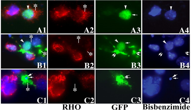
Figure 5.
The expression of mutant PRPF31 significantly inhibited the expression of rhodopsin protein. After the transfection of murine primary retinal cells with plasmids expressing either wild-type or mutant PRPF31 as GFP-fusion proteins, wild-type (A1-A4), N371 (B1-B4), or N256 (C1-C4) PRPF31 proteins were detected in the transfected primary retinal cells by monitoring GFP fluorescence (A3-C3), and rhodopsin expression was demonstrated by immunofluorescent staining (red fluorescence) using anti-rhodopsin antibody followed by staining with the secondary antibody conjugated with Cy3 (A2-C2). The nuclear morphology was revealed by staining with bis-benzamide (A4-C4). A1-C1 show the superimposed images in columns 2-4. Single arrowheads or arrows indicate nuclei with normal morphology. The cells marked with double arrows show abnormally condensed nuclei or dissolution of nuclei, as shown by bis-benzimide staining. Rhodopsin staining signal shows a significant reduction in cells expressing mutant PRPF31 proteins compared with those expressing the wild-type PRPF31 (compare cells marked by an asterisk in A with those in B and C; also compare transfected cells with neighboring nontransfected cells).

